| Zeit: | 15. Oktober 2022, 09:00 – 16:30 Uhr |
|---|---|
| Download als iCal: |
|
Program (more Details in the next weeks)
9:00 Prof. Dr.-Ing. Oliver Sawodny, Speaker of the RTG 2543; University of Stuttgart; Institute of System Dynamics
The welcoming will be done by Prof Stenzl on behalf of Prof. Sawodny
"Welcome"
9:20 Prof. Dr. Ute Neugebauer; University of Jena, Germany; Institute of Physical Chemistry, Clinical Spectroscopic Diagnosis
"Concepts for Intraoperative Localization and Tissue Differentiation"
Abstract: The intraoperative information gain is embedded in a higher-level pre- and post-operative learning loop that is intended to continuously improve the reliability of the conclusion. In order to exploit the information content of multiple sensor channels, sensor measurements must be provided with consistent local information. Finally, data-driven methods can be used to realize multisensor data fusion for intraoperative tissue differentiation. In this talk, concepts for intraoperative localization of sensor measurements, as well as data-driven classification concepts will be presented. A focus will also be placed on the efficient use of data to include existing labeled databases in addition to acquired data from partnering projects.
This contribution will present the potential of label-free Raman imaging to precisely localize and characterize intracellular pathogens directly inside intact host cells. Quantitative information about the metabolic phenotype can be extracted from 3D false color Raman images without any additional labelling technique and reveal important information on the life cycle of intracellular pathogens. Exemplarily, this will be shown for the obligate intracellular pathogen Coxiella burnetii, the active agent of the Q-fever, as well as for the facultative intracellular pathogen Staphylococcus aureus.
S. aureus is also one of the most frequent causes of sepsis and is involved in difficult-to-treat invasive tissue infections, such as osteomyelitis. Here, it will be presented how fluorescence and Raman imaging can be utilized to gain information of pathogen localization in tissue slices from a chronic osteomyelitis mouse model.
10:05 Presentation of the Research Focus A: Sensordevelopment
Valese Aslani1, Felix Fischer1, Lucas Becker3, Paul Kalwa4, Carina Veil2
1 Institute of Applied Optics, University of Stuttgart, 70569 Stuttgart, Germany
2 Institute for System Dynamics, University of Stuttgart, 70563 Stuttgart, Germany
3 Department for Medical Technologies and Regenerative Medicine, Institute of Biomedical Engineering,
University of Tübingen, 72076 Tübingen, Germany
4 Institute of Applied Physics, University of Tübingen, Pfaffenwaldring 9, 72076 Tübingen
"Sensor Concepts for Tissue Discrimination of Urological Specimens"
Abstract: Multiple institutes from the universities of Tübingen and Stuttgart with different core competencies joined forces to tackle the same problem of tissue differentiation. Within the last 2.5 years, five sensor concepts emerged from the fields of elastomechanics, optics, and electrodynamics. The two proposed mechanical sensors imitate palpational examinations by physicians and translate structural changes into digital information. The other three concepts deal with spectral changes in optical and electrical properties. All five sensors are validated using urological specimens of tumor and urothelia provided by the university hospital in Tübingen. In the long run, the concepts should support and complement each other to provide the best possible predictions for the examined tissue.
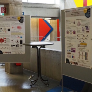
10:35 Coffee-Break, Poster Presentation of the RTG 2543
10:50 Prof. Dr. Hans Kreipe; MHH Hannover; Germany, Institute for Pathology
"Determination of preoperative therapy response for optimization of risk-adapted cancer therapy"
Abstract: Ki67 represents an immunohistochemical nuclear localized marker that is widely used in surgical pathology. Nuclear immunoreactivity for Ki67 indicates that cells are cycling and are in G1- to S-phase. The percentage of Ki67-positive tumor cells (Ki67 index) therefore provides an estimate of the growth fraction in tumor specimens. In breast cancer (BC), tumor cell proliferation rate is one of the most relevant prognostic markers and Ki67 is consequently helpful in prognostication similar to histological grading and mRNA profiling-based BC risk stratification. In BCs treated with short-term preoperative endocrine therapy, Ki67 dynamics enable distinguishing between endocrine sensitive and resistant tumors. Unlike triple-negative BC or the HER2-type of BC, which may exhibit dramatic response and will even completely vanish in a considerable proportion of cases during preoperative chemotherapy, luminal cancers, which make up the majority of breast cancer (75%) usually do not respond with major tumor shrinkage to endocrine therapy, and complete remission is rare. Whereas incomplete remission in response to preoperative therapy in HER2-positive or triple-negative cancers indicates at least partial resistance to therapy in the non-luminal types of BC, resistance to endocrine therapy providing the mainstay of treatment in luminal BC does not become clinically manifest until relapse will have occurred during treatment. As a possible indicator for endocrine sensitivity of luminal BC Ki67 has been proposed. In order to guide systemic therapy in early BC by Ki67-determined proliferative response to short-term 3-week preoperative endocrine therapy the multicentric, prospective, randomized ADAPT trial on more than 5.000 BC patients was conducted. In N0-1 recurrence score 12-25 patients with age <50years and treated with endocrine therapy alone, outcome of endocrine responders (⩽10% Ki67 after 3weeks) was superior when compared to non-responders. Thus, omission of chemotherapy in early BC of premenopausal patients with limited nodal burden and intermediate RS can be based on an easy accessible prognostic marker, provided by Ki67 response to short-term endocrine therapy. Missing Ki67 response to short-term endocrine preoperative therapy was associated with genetic aberrations, potentially conferring endocrine resistance like TP53 mutation, which has also been found in more than 25% of relapsing luminal BCs under therapy. Assessment of preoperative therapy response in cancer provides a novel tool with a bunch of benefits for patients. Reduction of tumor size limits the need for extensive surgical resections. In addition, this approach yields information about therapy response and resistance with the chance to reduce chemotherapy in responsive cases, and to address specifically therapy resistance with novel prospective trials.
11:35 Presentation of the Research Focus B: Modeling & Classification
Johannes Schüle1, Simon Holdenried-Krafft2, Peter Somers1
1 Institute for System Dynamics, University of Stuttgart, 70563 Stuttgart, Germany
2 Department of Computer Science, University of Tübingen, 72076 Tübingen, Germany
"Concepts for Intraoperative Localization and Tissue Differentiation"
Abstract: The intraoperative information gain is embedded in a higher-level pre- and post-operative learning loop that is intended to continuously improve the reliability of the conclusion. In order to exploit the information content of multiple sensor channels, sensor measurements must be provided with consistent local information. Finally, data-driven methods can be used to realize multisensor data fusion for intraoperative tissue differentiation. In this talk, concepts for intraoperative localization of sensor measurements, as well as data-driven classification concepts will be presented. A focus will also be placed on the efficient use of data to include existing labeled databases in addition to acquired data from partnering projects.
12:05 Prof. Dr.-Ing. Ulrike Thomas; University of Technology Chemnitz; Germany, Robotics and Human-Machine-Interaction Lab
"From Manipulation Skills to Telemanipulation for Medical Applications"
12:50 Lunch Break, Poster Presentation of the RTG 2543
14:00 Assistant-Prof. Dr. Julia Fleck, Center for Biomedical and Healthcare Engineering, Saint-Etienne, France
"Mathematical frameworks for integrating and modeling biomedical data"
Abstract: Mathematical data-driven approaches are especially suited for investigating the dynamics of complex diseases, such as cancer, and designing optimized treatment protocols for such diseases. I will introduce a mathematical programming formulation that integrates data to model the temporal order of cellular events, and illustrate its application to a pancreatic cancer dataset. I will also present algorithms to quantify and analyze the outcomes of different therapies, and exemplify their use through a case study of personalized therapy design for advanced prostate cancer.
14:45 Presentation of the Research Focus C: Surgery and Pathology
C1: Dr. Simon Walz (Universitätsklinik für Urologie, Tübingen)
"Optimizing culture media for Bladder cancer organoids"
C2: Dr. Lea Volmer (Universitätsklinikum Tübingen, Department für Frauengesundheit)
"Tissue differentiation in gynecology with a focus on the peritumoral milieu and micrometastasis"
C3: Dr. Massimo Granai/ Dr. Yvonne Montes (Institut für Pathologie und Neuropatholgie, Universität Tübingen)
"Development of a multiparametric tissue classification for tumors using histology as the gold standard".
Multiparametric analysis of ductal invasive breast cancer (NST) using targeted sequencing, image analysis and deep learning algorithms
Tilman Schotte, Massimo Granai, Ivonne Montes-Mojarro, Irina Bonzheim, Simon Holdenried-Krafft, Lea Volmer, Hendrik Lensch, Sara Y. Brucker, Annette Staebler and Falko Fend.
Abstract: With about 80-90% of all breast cancer cases, the invasive ductal carcinoma of no special type (IDC NST) is the most frequent subtype. Due to the high number of cases every year the interest in improving its grading and classification is very high. To this day the TNM-classification and Elston - Ellis grading is the gold standard to classify breast cancer. In addition, a molecular classification including the analysis of hormonal receptors and the proliferation marker Ki67 was established in the recent past. Although such methods improved the patient risk stratification there are still inaccuracies. That is particularly true for G2 tumors, in which such a grey zone may strongly impact on the therapeutic decision making. To further improve the grading of IDC NST we aim to establish a multiparametric approach to analyse whole slide specimens. This includes the analysis of the mitosis marker pHH3 and proliferation marker Ki67 in both resection specimens and core biopsies. In addition to the visual evaluation by trained pathologists we use the open-source digital analysis tool QuPath to eliminate interobserver biases. Moreover, our aim is to integrate the phenotypical results with molecular and genetic investigations by applying targeted NGS for p53 and ProSigna gene expression. Our study includes a learning cohort of 72 cases. Furthermore, we collected a larger validation cohort of 120 cases with a follow-up of a minimum of 10 years to confirm these results. Preliminary results show that the QuPath analysis of Ki67 matches the visual results in the vast majority of the cases. On the other hand, the results of mitosis scoring with pHH3 staining through automatic analysis are higher than those performed visually. To further confirm our findings other studies on larger collectives are needed.
Multiparametrical classification of muscle invasive bladder cancer using immunohistochemistry, targeted sequencing and image analysis
Sina Staehle, Ivonne Montes-Mojarro, Massimo Granai, Irina Bonzheim, Simon Walz, SIimon Holdenried-Krafft, Arnulf Stenzl, Hans Bösmüller and Falko Fend.
Abstract: 25% of bladder tumors are muscle-invasive by the time of diagnosis. New molecular classifications strongly suggest recognizing histological subtypes as predictors of aggressive tumor behavior. The main objective of this work is to develop a multiparametric classification for muscle invasive bladder cancer (MIBC), by using image analysis, immunohistochemical stains for HER-2, FGFR3, CK5/6, Mib-1, p40 and Cyclin D1 and targeted sequencing including a panel with 65 genes. We will review and analyze the clinical and histological data of 132 patients diagnosed with either papillary muscle invasive bladder cancer or muscle-invasive histological subtypes. So far, the immunostaining profile including HER2, CK5/6, cyclin D1, and p40 demonstrated differential expression in papillary MIBC and its variants (P<0.05). However, the proliferation rate (Mib-1) and FGFR3 showed comparable levels of expression. Therefore, we aim using different molecular parameters to look for specific clusters and thereby propose a new multiparametric classification of MIBC. Further validation of this classification and its biological and clinical implication will be the subject of further studies.
15:15 Coffee-Break, Poster Presentation of the RTG 2543
15:30 Prof. Dr. Andreas Kolb; University of Siegen, Germany, Lehrstuhl für Computergrafik
"Interactive Scene Reconstruction – Fundamentals and New Trends"
Abstract: The advent of affordable consumer grade RGB‐D cameras has brought about a profound advancement of visual scene reconstruction methods. Both computer graphics and computer vision researchers spend significant effort to develop entirely new algorithms to capture comprehensive shape and appearance of models. Still, there is no overall optimum regarding performance along relevant dimensions, being it high reconstruction quality or real‐time performance on commodity hardware.
The first part of this talk will give an overview of fundamental approaches, data structures and techniques applied in the field of interactive scene reconstruction and common challenges under research in this field. In the second part new trends in approaching the challenges of interactive scene reconstruction are discussed.
16:15 Prof. Dr.-med. Arnulf Stenzl, Deputy-Speaker of the RTG 2543; Eberhard-Karls-University Tübingen, University Hospital, Department of Urology, Germany
"Summary"
16:30 End of Symposium


