Tissue Elasticity as Diagnostic Marker
Elasticity is a crucial parameter of living cells and tissue and one of the parameters that are changing during tumor proliferation. Hence there are currently different approaches to make tissue elasticity a diagnostic marker in medicine. So far, ultrasound elastography and magnetic resonance elastography showed that it is possible to identify tumor tissue through its elastic parameters. Therefore, further approaches were developed to measure elasticity of human tissue in vivo, e.g., using torsion-resonance or aspiration. Also, optical approaches like coherence tomography or confocal microscopy (see project A1) are being pursued. However, complex instrumental setups are necessary for all of these known procedures and approaches.

Water Jet Elastography
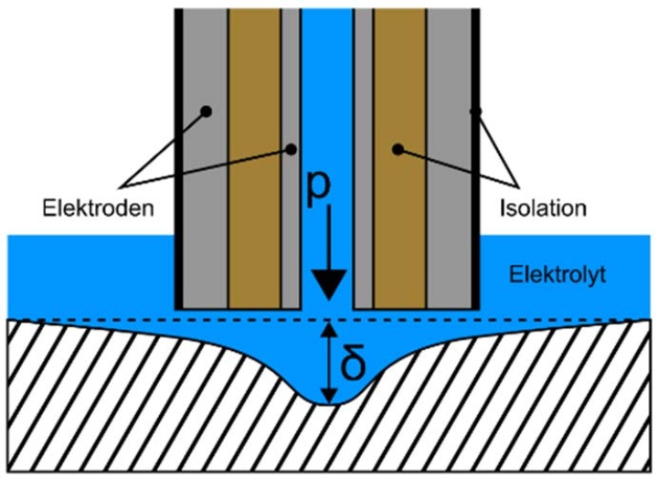
In this project a new application in form of water jet elastography should be investigated and a new sensor should be developed. The underlying principle of the sensor is based on the deformation of tissue due to a water jet (see figure). This water jet is applied through a nozzle above the tissue and indents the tissue elastically. The local indentation depth is related to the mechanical properties of the tissue: for softer tissue, the indentation depth increases and therefore the electrical impedance between the two electrodes decreases. This allows the determination of the elasticity of the tissue. To ensure a sufficient conductance and biocompatibility, a saline solution is used.
Part of this project is therefore the adaption of the principle of water jet elastography on tissue in the magnitude of millimeters. In comparison to other methods of elasticity measurement of tissue the principle of water jet elastography is quite simple and its application is relatively inexpensive, which is beneficial for clinical use.

Emily Hellwich
M. Sc.PhD Student A4
[Image: Emily Hellwich]

Tilman Schäffer
Prof. Dr.Principle Investigator of Subproject A4
[Image: Tilman Schäffer, Universität Tübingen]


